This reflected energy indicate is found by theprobe. Enough time it requires for the indicate to travel back once again to the probe is digitally tested and applied to determine, along with to produce, the level of the tissue. The frequencies useful for medical ultrasonography are generally in the product range of 1 to 15 MHz. Larger frequencies have a lowered wavelength, making photographs with a greater resolution. However, the reduced total of the trend is increased at larger frequencies, so in order to most efficiently enter deeper tissues, a lesser volume (3-5MHz) is used.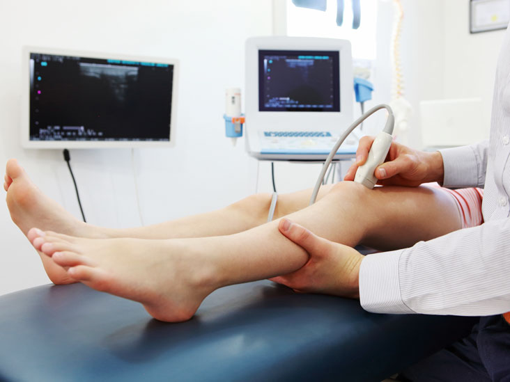
Ultrasound transducers make photos of muscle and delicate structure, useful for defining boundaries between stable and fluid-filled spaces. Ultrasound transducers give live photographs, enabling operators to pick the most readily useful areas for rapid diagnoses. They display the structure and function of inner organs. It give a good solution to study the musculoskeletal system to detect difficulties with muscles, structures, tendons and joints. It also assist in determining obstructions, stenosis and other general abnormalities.
Contemporary, high-class ultrasoundsystems utilize the most readily useful 4d ultrasound werribee transducers engineering, in conjunction with excellent processors and an easy to use interface. The picture quality depends mostly on the ultrasound transducer, which can be leading conclusion that sends and gets the mechanical energy. Contemporary ultrasound transducers provide consumers unparalled and unparalleled multi-modality ultrasound experience.
Carotid ultrasonography, applied to evaluate blood movement in to the carotid arteries, as well as the intra-cerebral arteries. Echocardiography, that will be an ultrasound that reveals the action of the center, while the muscle dilates and contracts. Emergency medical professionals frequently make use of a form of probe. Also, it is used in the ER as a routine way of easily assessing the reason for a patient’s abdominal pain.
Urologists often use ultrasound transducers to find the degree of water that’s in a patient’s bladder. Gynecologists might perform a pelvic sonogram using an ultrasound to see a graphic of the pelvic ground in women and identify any abnormalities. More descriptive photos of the tendons, nerves, muscles, ligaments and different delicate muscle places that could have been suffering from harm or trauma. Arterial probe is utilized by cardiologists to test for possible obstructions in the arteries, or to identify DVT. In gastroenterology, health practitioners can see the abdomen utilizing an probe to view organs including the aorta, pancreas, gall bladder, kidneys, liver and spleen.
You can find four types of available ultrasound image. The decision that type of picture to use is dependent upon the objectives for a specific check, the phenomena being investigated and what equipment is available. The most typical and kind of ultrasound picture is a series of level, two-dimensional mix part photos of the scanned tissue. Known merely as 2d ultrasound, that method of checking is still standard for many diagnostic and obstetric scenarios after having a half-century of use.
Recently, second photos have been expected into three-dimensional representations. This is attained by checking muscle mix parts at a variety of perspectives and reconstructing the data obtained into a three-dimensional image. A typical use for 3d ultrasound pictures is to offer a far more complete and reasonable picture of a building fetus. By upgrading 3d ultrasound photos in quick series, sonographers may also create 4d ultrasound pictures. In the 4d ultrasound, the next aspect, time, provides motion and generates the absolute most reasonable illustration of all.
In some cases, 3d and 4d ultrasound pictures may possibly reveal abnormalities perhaps not easily seen using 2d ultrasound. For pregnant parents and nearest and dearest, the capacity to see reasonable pictures of an unborn baby in the womb may be worthwhile and heartwarming although the medical neighborhood in general cautions against performing ultrasound checks exclusively because of this purpose.